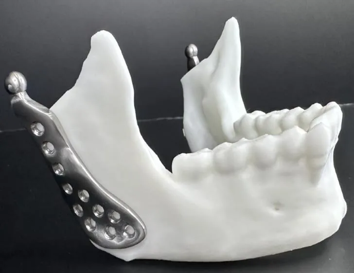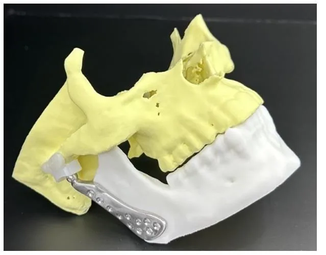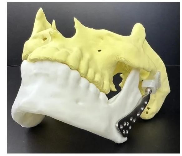

Case 2: Bilateral TMJ Implant
Tanı: Custom bilateral TMJ implant treatment was planned for the patient diagnosed with ankylosing spondylitis.
Ankylosing Spondylitis TMJ Implant
Patient-specific bilateral TMJ implant treatment is planned.
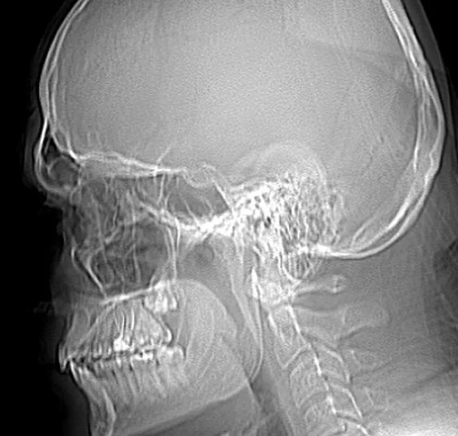
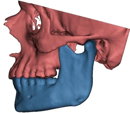
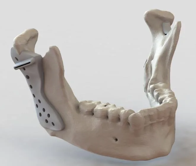
Anatomical bone models were created from the patient's DICOM data.
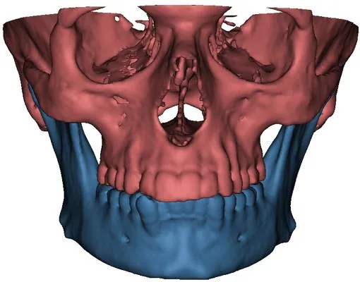
The maxilla and mandible were segmented by adjusting the Hounsfield unit thresholds in the CT images for the individual patient.
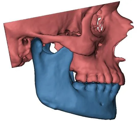
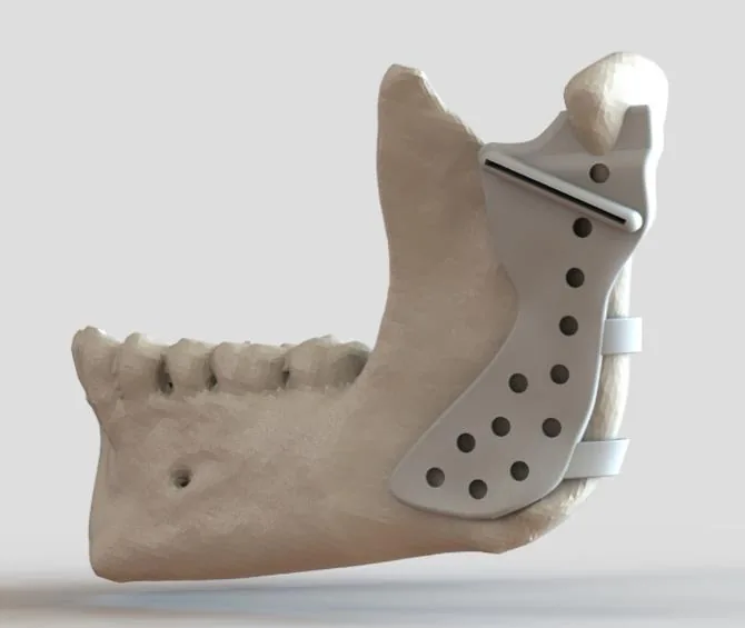
Implant designs were created without moving the mandible (preserving anatomical occlusion).
Bilateral TMJ implant components, designed according to patient-specific anatomical morphology and relevant physician requests, were obtained.
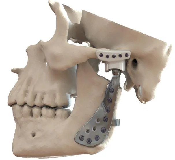
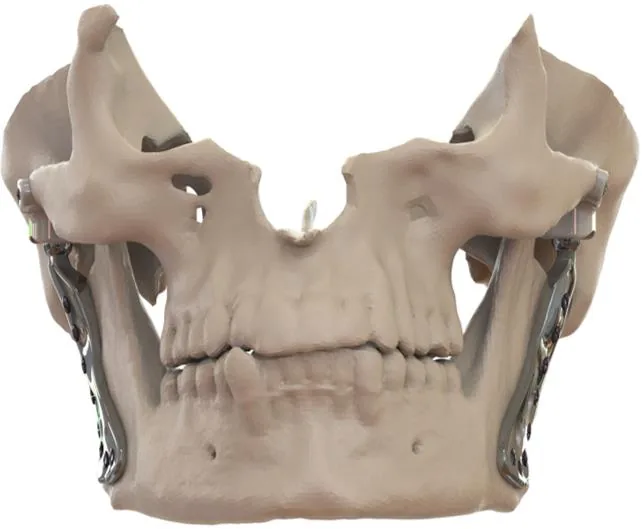
Before implant assembly, the articular tubercle (AT) joint was adjusted to increase the fit of the fossa prosthesis.
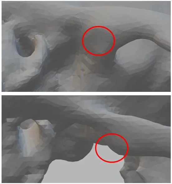
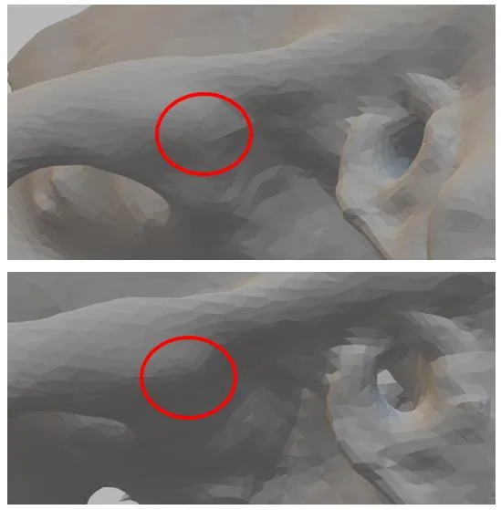
Special Guides
Custom mounting and cutting guides.
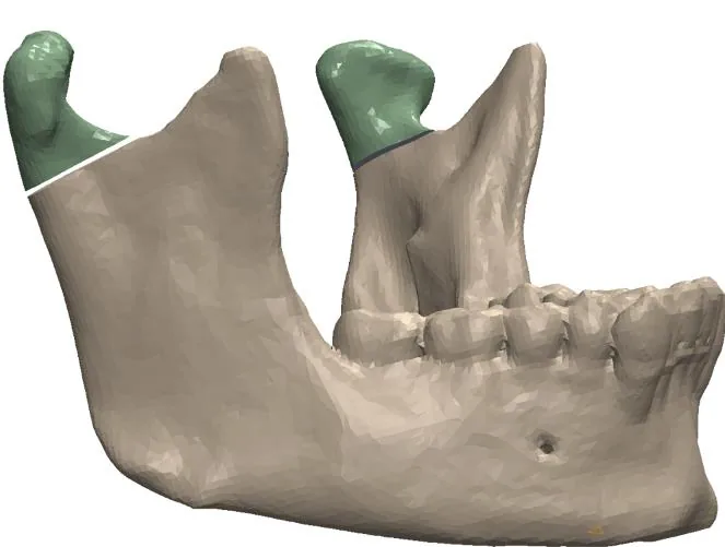
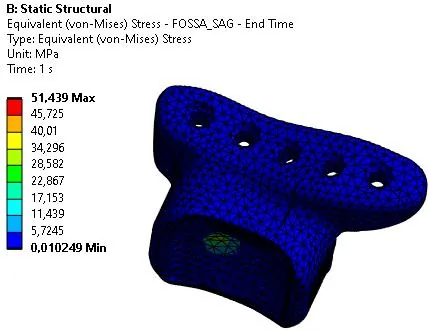
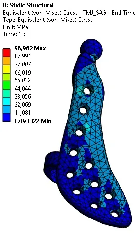
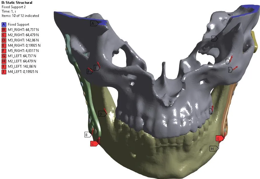
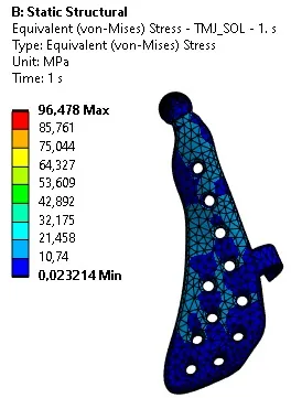
Finite Element Analysis

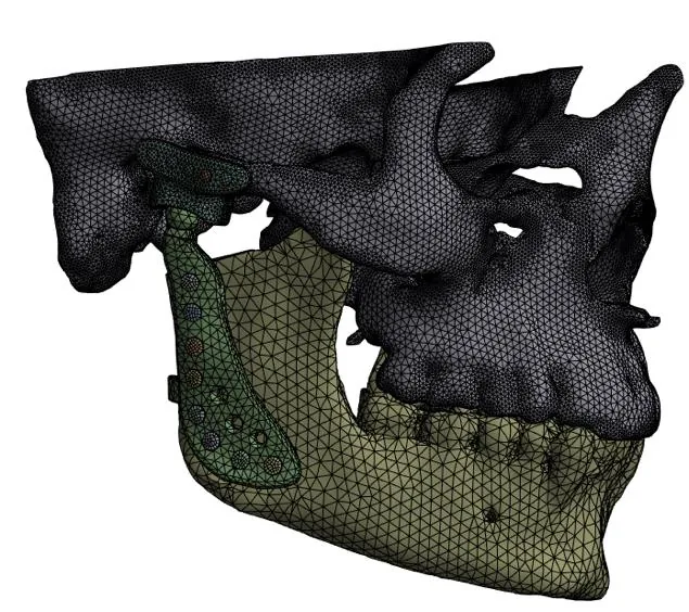
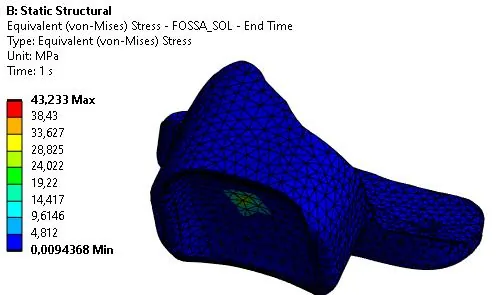
1- Finite element analysis of the designed components was performed in ANSYS software.
2- The mechanical properties of Medical Ti6Al4V-Eli and Medical UHMWPE materials were obtained from the manufacturer, and those of the bone structures from the literature.
3- Analyses of the meshed model were performed under specified boundary conditions (considering muscle forces) prior to analysis.
4- As a result of the analysis, average stresses of 45 MPa in UHMWPE components and 100 MPa in Ti6Al4V components, excluding local peak values, were determined.
5- Local peak values are related to geometric irregularities caused by plunging in bone morphology.
Production and Post Process
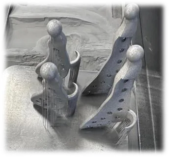
Additive manufacturing of metal components
Implants were produced using Selective Laser Melting (SLM) method with medical Ti6Al4V-Eli alloy powder.
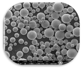
The SEM image of the powder used shows its sphericity.
The following parameters were used in production:
- Laser power: 80W
- Scanning speed: 1125mm/s
- Layer thickness: 25µm
- Hatch spacing: 45µm
CNC manufacturing of UHMWPE components
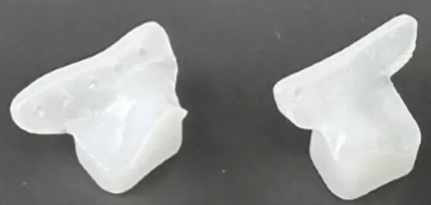
Medical UHMWPE components were produced in a CNC 3-Axis Vertical Machining Center.
Different holders were designed and produced for the manufacturing of the components.
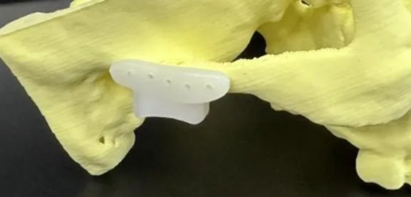
After production, preliminary checks were performed on bone models produced with the FDM method.
Post process operations
The implants were polished using grit 240, 400, 600 and 1200 SiC sandpapers respectively, and finally polished with a-alumina powder with a particle diameter of 0.05 µm.
After polishing, it was washed ultrasonically in alcohol at 53KHz frequency for 15 minutes at 26°C.